


Sonography is necessary during different stages of pregnancy. During the second trimester of the pregnancy; between 18 to 20 weeks an ultrasound determines that things are moving ahead as they should. When you decide to have a sonography, we suggest you connect with Sunrise Diagnostic team because, it is one of the best sonography center in Pune. We are sure you would recommend it for full body health checkups to your friends and family.
Apart from the fact that a sonography offers patients those special moments of the first picture of the baby, the ultrasound or sonography has some important diagnostic functions. They provide critical information of the mother and the health of the baby.
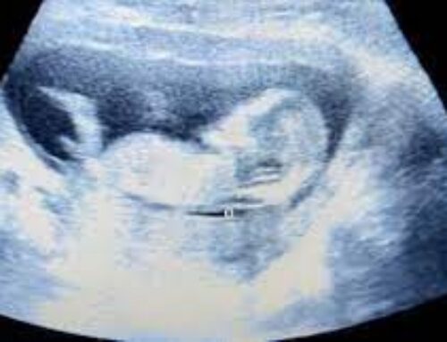
Sonography is utilized to analyze and monitor the developing fetus, the uterus and placenta. This is necessary to ensure the normal development of fetus and placenta.
Basically, ultrasound imaging is utilized to assess development during pregnancy in the following manner:
Table of Contents
Confirm the pregnancy
1. Gestational Age:
40 weeks gestation is considered as normal pregnancy but, in medical terms it’s considered from 37 to 41 weeks.
Why is it important to verify the gestational age of a developing fetus?
The baby’s growth is compared with well established growth charts. This is to ensure if the baby is growing normally.
Gestational age is arrived at after noting the dates informed by the mother about her last menstrual period so that the due date can be confirmed. This way the doctor ensures that the baby isn’t delivered way to early or too late.
2. Issues with the placenta:
During pregnancy the placenta’s position in the uterus is significant for the health of the baby and the mother. An ultrasound can identify the following issues:
Vasa Previa:
It is a condition where the baby’s blood vessels run near the cervix. Due to this, vessels might get raptured in case the mother’s membrane raptures.
In case of vasa previa the patient has to hospitalized during the pregnancy and in case of a cesarean section.
Placenta Previa:
When the placenta lies usually low in the uterus, either it’s next to or covers the cervix, it is referred as placenta previa. It’s common to have the placenta lying close to the opening of the cervix in the early stages of the pregnancy. It can also move up the uterine wall as the baby grows.
But if it’s low at the time of labor then it’s a bit troublesome since it can lead to catastrophic bleeding that can injure the mother and the baby too. How will an ultrasound help?
It will determine if the situation needs a cesarean section delivery.
Placenta Percreta:
In this condition the placenta is grown through the uterine wall.
Placenta Accreta:
The blood vessels and other placenta parts grow deep into the uterine wall. This causes problem as the placenta has to detach from the uterine wall after the child is born. While in placenta accrete, part or all the placenta has a grip on it.
This can be a problem because the placenta normally detaches from the uterine wall after childbirth. With placenta accreta, part or all of the placenta remains firmly attached. This can cause severe blood loss after delivery.
Placenta Increta:
In this condition the placenta invades the uterus muscles.
3. Keeping a check on the congenital anomalies:
Most parents wish to ensure that the baby is healthy and has no genetic problems or genetic issues. They can choose to terminate the pregnancy if they know about it. Alternatively, they can be prepared for the difficulties that they need to face.
4. Multiple pregnancies:
If the pregnancy includes multiple babies, it carries some risks and must be monitored regularly. One such complication can be twin to twin transfusion.
5. Monitor Fetal Growth:
When the baby’s growth is slow as compared to the norms then it indicates problems with the placenta or health of the baby. Early intervention is always better in all cases.
6. Monitor Fetal Position
It is important to know the placement and position of the baby as it affects the method of delivery.
7. Monitor the Level of Amniotic Fluid:
Fetus produces amniotic fluid; its either too much or too little fluid that indicates pregnancy problems that require early intervention.
Other tests can be done easily when its preceded and guided by an ultrasound.
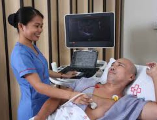
What are the different types of ultrasounds?
- Transvaginal ultrasound is done through the vagina. A thin transducer shaped is put into the vagina. You might feel a bit of a pressure while doing it but causes no pain. This takes 20 minutes and your bladder has to be full during this procedure.
- 3-D ultrasound takes many pictures at once. It takes 3-D image that has immense clarity. This type of ultrasound is used to ensure that the baby’s organs are developing and growing normally. It also keeps a check on the problems in the uterus and abnormalities in the baby’s face.
- 4- D ultrasound is like the 3-D ultrasound and pictures the baby’s movement on a video.
- Fetal echocardiography uses sound waves to keep a check on the heart of your baby while developing. Fetal echo finds out the health defects even before birth of the baby.
We believe that your curiosity has been satiated after reading this blog post. Visit our website: www.sunrisediagnosis.com and blog: www.sunrisediagnosis.com/blog to know more about different diagnostic and imaging tests. Infact, Sunrise Diagnostic has the best health checkup packages in Pune. Be rest assured that you are in safe hands because, we are one of the best diagnostic center in Pune.

